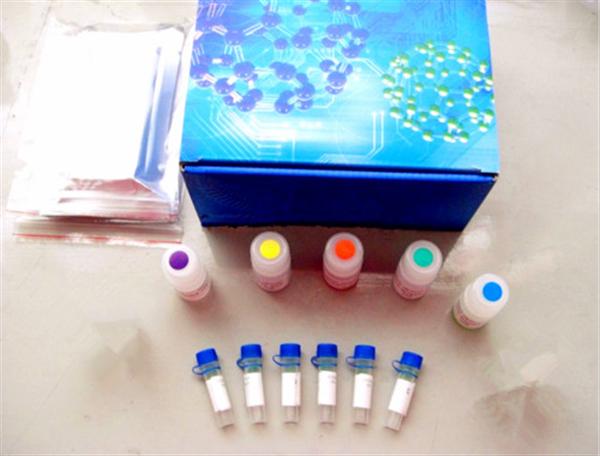
The most common method for detecting antigens in an ELISA kit is the sandwich ELISA technique. This approach is widely used in clinical and research settings due to its high sensitivity and specificity.
Here are the key steps involved:
- A specific antibody is immobilized onto a solid-phase carrier, such as a microplate well. After incubation, unbound antibodies are washed away, leaving only the bound antibodies on the surface.
- The sample containing the target antigen is added and allowed to incubate. The antigen binds to the immobilized antibody, forming a solid-phase antigen-antibody complex. Any unbound substances are then removed through washing.
- An enzyme-conjugated secondary antibody is introduced. This antibody recognizes and binds to the antigen, forming a complete "sandwich" structure. Unbound conjugates are thoroughly washed away.
- A substrate is added, which reacts with the enzyme to produce a detectable color change. The intensity of the color is proportional to the amount of antigen present in the sample, and this is measured using a spectrophotometer.

This method is particularly effective for detecting macromolecular antigens such as proteins like HBsAg, HBeAg, AFP, hCG, and others. To set up the assay, it's essential to have a suitable antibody against the target antigen. If using polyclonal antisera, it's preferable to use antibodies from different species for coating and labeling to avoid cross-reactivity. For monoclonal antibodies, two antibodies that recognize different epitopes on the antigen are typically used—one for coating the solid phase and the other for enzymatic labeling.
The two-site sandwich method offers high specificity and allows for one-step detection, where the specimen and enzyme-labeled antibody can be incubated together. However, there is a potential issue known as the "hook effect," which occurs when the concentration of the antigen is too high. In such cases, excess antigen may bind to both the solid-phase and enzyme-labeled antibodies, preventing the formation of the sandwich complex. This leads to a decrease in signal, resulting in an underestimation of the actual antigen concentration.
The hook effect is visually represented by a curved standard curve, resembling a hook after reaching its peak. In severe cases, this might even lead to false-negative results. Therefore, when testing samples with high antigen levels—such as HBsAg, AFP, or urine hCG—it’s important to be aware of the upper limit of the measurable range. Using high-affinity monoclonal antibodies can help reduce the hook effect and improve accuracy.
If the antigen has multiple identical epitopes (such as the 'a' determinant of HBsAg), a single monoclonal antibody can be used for both coating and labeling. However, in the case of HBsAg, it's crucial to consider the presence of different subtypes (adr, adw, ayr, ayw). While these subtypes share the same 'a' determinant, they may react differently depending on the monoclonal antibody used. This is especially important when using the sandwich method for detection.
The double antibody sandwich method works best for antigens with at least two binding sites (divalent or higher). It is not suitable for small molecules or haptens, which lack sufficient epitopes to form a stable sandwich complex.
Ejuice Vg Pg,Concentrate,Disposable Vape Pen,Wholesale Vape,Oem Odm Vape,Pouch Vape Pen,Pouch Ecig
Niimoo Innovative (HK) CO., LIMITED , https://www.niimootech.com